
Throughout the past 30 years, scientists have been extensively researching organisms that have the ability to produce the ferromagnetic mineral magnetite. Magnetite is a black mineral form of iron oxide that crystallizes in the cubic or isometric system, namely all crystals which have their crystallographic axes of equal length at 90 degrees to each other. It is a mixed Iron (II) Iron (III) oxide, Fe3O4, and is one of the major ores of iron that is strongly magnetic. Some varieties, known as lodestone, are natural magnets; these were used as compasses in the ancient world.
The discovery of a biogenic material (that is, one formed by a biological organism) with ferromagnetic properties and found to be magnetite was the first breakthrough toward an understanding as to why some animals have the ability to detect the earth’s magnetic field. Searches for biogenic magnetite in human tissues had not been conclusive until the beginning of the 1990’s when work with high-resolution transmission electron microscopy and electron diffraction on human brain tissue extracts of the cerebral cortex, cerebellum, and meninges (membranes surrounding the brain and spinal cord) identified magnetite-maghemite crystals.
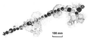
Magnetite Crystals
Magnetite Crystals under Low Magnification
These magnetite crystals were found to be organized into linear, membrane-bound chains a few micrometers in length, with up to 80 crystals per chain. Furthermore individual crystals have their {111} aligned along the length of the chain axes (the “easy” direction of magnetization). The {111} crystal alignment has been interpreted as a biological mechanism for maximizing the magnetic moment per particle, as the {111} direction yields approximately 3% higher saturation magnetization than do other directions. This prismatic particle shape is also uncommon in geological magnetite crystals of this size, which are usually octahedra. The crystal morphology was found to be cubo-octahedral with the {111} faces of adjacent crystals lying perpendicular to the chain axis.
All the magnetite crystals that have been examined to date are single magnetic domains, which means that they are uniformly and stably magnetized and have the maximum magnetic moment per unit volume possible for magnetite. Elemental analysis, by energy-dispersive X-ray analysis, electron diffraction patterns, and high resolution transmission electron microscopy lattice images, showed that many of the particles were structurally well-ordered and crystallographically single-domain magnetite. This means that the production of this biomineral must be under precise biological control.
Ferromagnetic crystals interact more than a million times more strongly with external magnetic fields than do diamagnetic or paramagnetic materials (deoxyhemoglobin, ferritin, and hemosiderin).With this finding researchers were posed with a fundamental question for biology, namely: What is the mechanism through which the weak geomagnetic fields are perceived by organisms that are able to precipitate crystals of a ferromagnetic mineral such as magnetite (Fe3O4)? Could these crystals use their motion in a variety of ways to transduce the geomagnetic field into signals that can be processed by the nervous system?
The presence of membrane-bound biomineral magnetite, which has been shown to have a biological origin, and the implication that some kind of mechanical coupling must take place between each compass magnetite particle and a mechanoreceptor, or at least a functionally equivalent mechanism allowing the position of the particle to be monitored by a sensory organelle in the body, is unique. Research has also found that the magnetite is produced by the cells of the organism when needed. Forms of advanced physical intelligence can directly tap into this information if they have a crystalline network within their brain cavity.
Scientists are now asking the fundamental question: What is magnetite doing in the human brain? In magnetite-containing bacteria, the answer is simple: Magnetite crystals turn the bacteria into swimming needles that orient with respect to the earth’s magnetic fields. Magnetite has also been found in animals that navigate by compass direction, such as bees, birds, and fish, but scientists do not know why the magnetite is present in humans, only that it is there.
We have also seen in research done in the late 1980s that proteins, DNA, and transforming DNA function as piezoelectric crystal lattice structures in nature. The piezoelectric effect refers to that property of matter which may convert electromagnetic oscillations to mechanical vibrations and vice versa. Studies with exogenously administered electromagnetic fields have shown that both transcription (RNA synthesis) and translation (protein synthesis) can be induced by electromagnetic fields and furthermore that direct current in bone will produce osteochondrogenesis (bone formation) and bacteriostasis, as well as affect adenosine triphosphate (ATP) generation, protein synthesis and membrane transport.

Single Magnetite Crystal in Human Brain
In the human brain, pyramidal cells are present and arranged in layers in the cortex of the two cerebra. The pyramidal cells act as electro-crystal cells immersed in extra-cellular tissue fluids, and seem to operate in the fashion of a liquid crystal oscillator in response to different light commands, or light pulses which, in turn, change the orientation of every molecule and atom within the body. Biogravitational encoded switches present in the brain allow a type of liquid network to release ions that induce currents to the surrounding coiled dendrites. Electron impulses from a neuron, on reaching the dendrite coil of the abutted cell, generate a micro amperage magnetic field, causing the ultra thin crystal, or liquid crystal in the pyramidal cell to be activated — in a very unusual way. On flexing, this ultra thin crystal becomes a piezoelectric oscillator, producing a circular polarized light pulse that travels throughout the body, or travels as a transverse photonic bundle of energy.
According to Einstein, matter is to be regarded itself as part, in fact the principle part, of the electromagnetic field, and electric energy is therefore the fundamental origin of our entire physical world. Consequently, in work published by The Academy For Future Science it has been cited that “under present biological conditions, evolutionary development in living bodies from earliest inception follows unicellular semiconductivity, as a living piezoelectric matrix, through stages which permit primitive basic tissues (glia, satellite and Schwann cells) to be supportive to the neurons in the human system where the primary source is electrical. This has been especially shown in bone growth response to mechanical stress and to fractures which have been demonstrated to have characteristics of control systems using electricity.”
Ongoing research has shown that bone has electrical properties. The bone matrix is a biphasic (two-part) semiconductor, i.e. a crystalline solid with an electrical conductivity. The collagen component of bone matrix is an N-type semiconductor and the apatite component a P-type. When tested for piezoelectricity, collagen turns out to be a piezoelectric generator while apatite is not. These function as two semiconductors, one an N-type, the other a P-type forming a PN-junction, which sets up a potential barrier and acts as an efficient rectifier, i.e. a semiconductor diode.
Mechanical stress on the bone thus produces a piezoelectrical signal from the collagen. The signal is biphasic, switching polarity with each stress-and-release. The signal is rectified by the PN-junction between apatite and collagen. The strength of the signal tells the bone cells how strong the stress is, and its polarity tells them what direction it comes from. Osteogenic (bone forming) cells, which have been shown to have a negative potential, would be stimulated to grow more bone, while those in the positive area would stop production of matrix and be resorbed when needed. If bone growth and resorption are part of one process, the electrical signal acts as an analog code to transfer information about stress to the cells and trigger the right response. Hence, stress is converted into an electrical signal.
An interesting property of PN-junctions of semiconductor diodes may be observed when current is run though the diode in forward bias, i.e. when there is a good current flow across the barrier. Some of the energy is turned into light and emitted from the surface and are therefore known as light-emitting diodes (LEDs). Researchers found that bone was an LED that required an outside source of light before an electric current would make it release its own light, and the light it emitted was at an infrared frequency invisible to us, but consistent.
With the use of an applied current of a few microamperes regeneration of the spinal cord, optic nerve and bone has been demonstrated and naturally generated electric currents have been linked to changes in developing embryos and in regenerating limbs.
During the past decades a great increase has taken place in research on the effects of non -ionizing electromagnetic radiation on biological systems. Much has been revealed about the human organisms on all levels but the question still being asked by scientists is: What electromagnetic signal might tune to a magnetic resonant energy which would alter the metabolic genetic regulation to bring about growth and repair? It has been considered by this author that tRNA molecules may play a central roll to cause cells to alter their normal properties which will then receive the original genetic transmission, given through a ‘spin point’ to a cell. These transmissions at the spin points, as discussed through research at The Academy For Future Science, may provide regenerating instruction for the manufacture of enzymes and proteins which are the building blocks for the ‘new tissue’ or the ‘new organ form’ which is regenerated on the physical plane. Projecting energy into the spin point allows for the formation of a blastema (mass of primitive type cells) that gives rise to the regenerated tissue. Thus, through the spin point, cells become the tissue responsible for the generation and transmission of direct current signals used in regeneration processes.
________________________________________
Definitions:
Paramagnetism is a weak magnetic condition of substances that have a positive but small susceptibility to magnetism.
Diamagnetism is the phenomenon exhibited by substances that are repelled by both poles of a magnet and thus lie across the magnet’s line of influence i.e. have a negative susceptibility to magnetism. All substances are diamagnetic.
Ferromagnetism is the phenomenon exhibited by substances such as iron that show increasing magnetization with applied magnetizing field and persists after the removal of the applied field.
Magnetic domain is one of the regions in a ferromagnetic solid in which all the atoms have their magnetic moments aligned in the same direction.
Crystal faces are represented by indices and when the indices are enclosed in braces, e.g. {111}, the indices refer to a complete group of faces.
________________________________________
References
Becker, R.O. and Selden, G. The Body Electric: Electromagnetism and the Foundation of Life. New York, NY: Quill, William Morrow, 1985.
Dubrov, A.P. The Geomagnetic Field and Life: Geo-magnetobiology. New York, NY: Plenum Press, 1978.
Hurtak, J.J. “The Power of Healing.” Lecture given to the members of the Bioenergetics Institute, Johannesburg, 1986.
Jacobson, J.I. “Exploring the potential of magneto-crystallization of genes and associated structures with respect to nerve regeneration and cancer.” International Journal of Neuroscience, 64 (1992):153-165.
Kirschvink, J.L. “Magnetite Biomineralization and Geomagnetic Sensitivity in Higher Animals: An Update and Recommendations for Future Study.” Bioelectromagnetics,10 (1989):239-259.
Kirschvink, J.L. et al. “Magnetite biomineralization in the human brain.” Proceedings of the National Academy of Sciences, 89 (1992):7683-7687.
Nordenstrom, B.W.W. “Impact of Biological Closed Electric Circuits (BCEC) on Structure and Function.” Integrative Physiological and Behavioral Science, 27 (1992):285-303.